

- European area (CE)
- Australia
- Mainland China
- Hong Kong, Macau
If it is for research purpose, it depends on the regulations of the specific region/country. Most of the region/country allows the use for research purpose.
Since 2013, Scolioscan® has been installed and used in more than 20 public hospitals, rehabilitation clinics and universities in Europe, Australia, Mainland China, Hong Kong and Macau. For details, please refer to the official website of Telefield Medical Imaging Limited and the case studies in our WeChat Official Account.
Telefield Medical Imaging Limited has a production site in Hong Kong and a production site in Changsha, China. The products produced from Hong Kong production site are sold to overseas countries, and the products produced from Changsha production site are sold to Mainland China.
The SCN800 series is ergonomically designed with supporting structures for the chest and hips, which help patients maintaining a natural standing posture during scanning. The system includes a 24-inch adjustable touchscreen monitor and has a large built-in data storage space, you do not need to prepare separated device by yourself when operating it.
Only the SCN800 series can support Scolio3D for 3D data analysis and the use of DICOM.
The portable design of the SCN200 series enables the possibility of scoliosis assessment in any setting. Unlike the stationary SCN800 series, the SCN200 series is ultra-light and portable, making scoliosis assessment more convenient and efficient. It weighs just 500 grams and can be stored in a custom carrying case.
The image resolution of the SCN200 system is slightly inferior to that of the SCN800 series, and is generally more suitable for large-scale scoliosis screening. At the same time, it can be used together with SCN800 series for further examination, progress monitoring and real-time treatment effectiveness.
It is suggested to use SCN800 series for professionals, this series consists of a spatial sensing system with the sensor embedded in the ultrasound probe. In addition to conventional B-mode imaging, it collects the spatial location of the vertebrae and forms the full spine image, together with Scolio3D for the full spinal profile analysis. This series provide a safe, cost-effective and accurate spinal assessments for professionals.
Professionals can choose SCN800 or SCN200 series. The SCN800 series is embedded with the most advanced ultrasonic module and spatial sensing system, which provides more flexibility. If the center or clinic has limited space for activities, you can choose the SCN200 portable series. The system has a net weight of 500 grams and palm-sized, allowing you to scan for scoliosis assessment at anytime and anywhere.
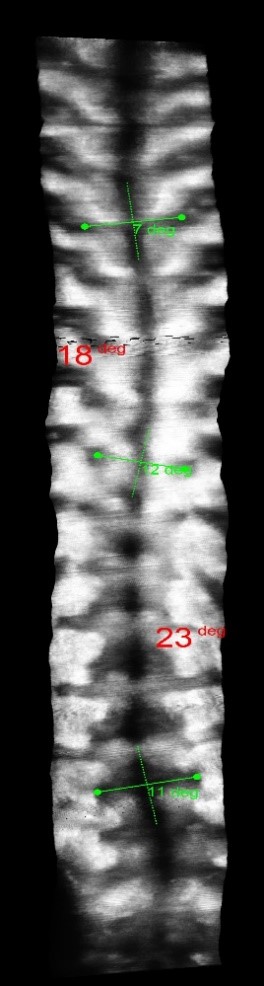
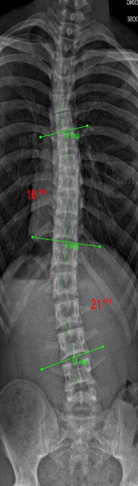
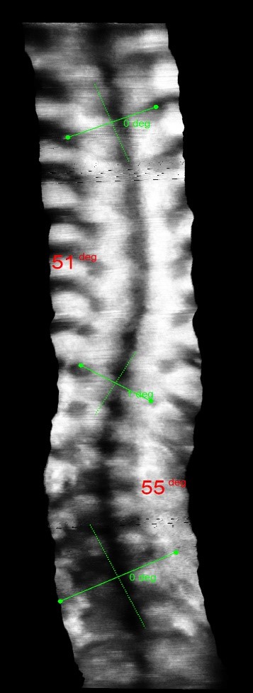
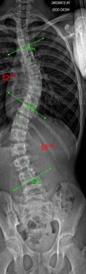
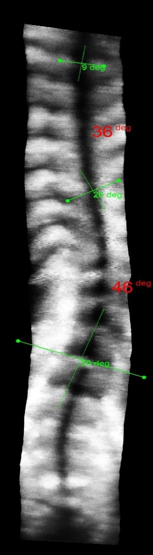
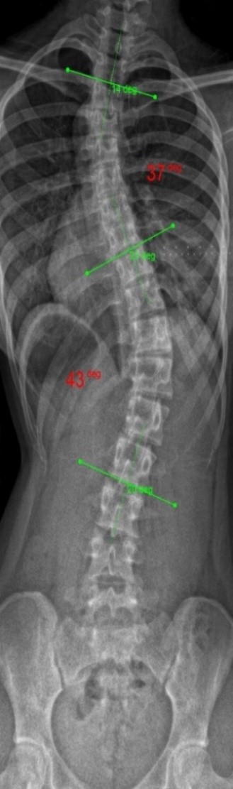
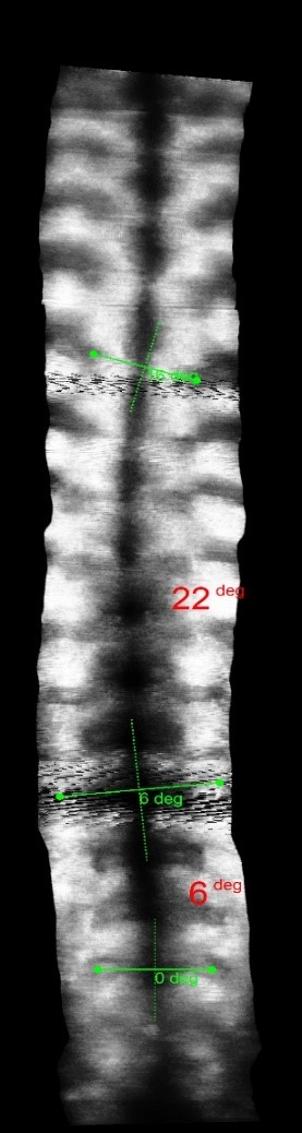
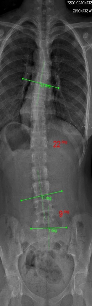
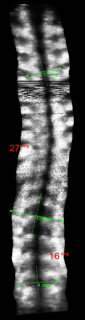
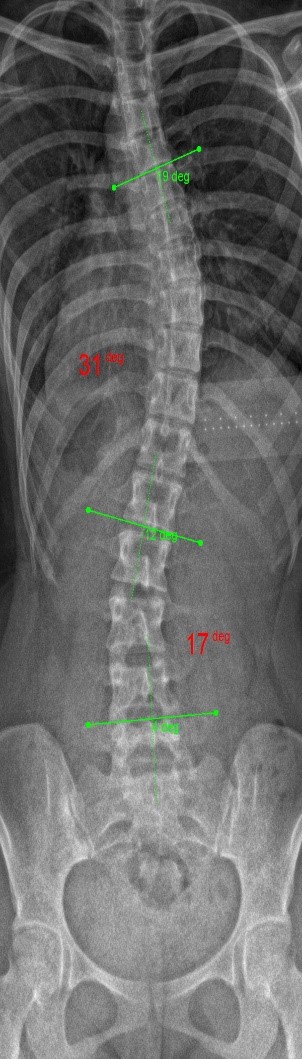
Scoliometer is based on the body surface curvature, and it provides only the body axial rotation angle during a bending test. Its error is large in comparison with Cobb angle. Scolioscan® uses the coronal image formed by 3D ultrasound scanning with the subject standing erectly, and it measures the lateral curvature of the spine, the same as X-ray Cobb angle.
X-rays are the current gold standard for scoliosis diagnosis, while MR is rarely used for quantitative detection of scoliosis. That is because MR requires the patient to be scanned in lying position, and it is time–consuming and expensive.
The clinical test results show that the SCN800 series is highly consistent with the X-ray, with a linear correlation of 0.94, similar results have also been published in SCI articles (Lee TT, Lai KK, Cheng JC, Castelein RM, Lam TP, Zheng YP. 3D ultrasound imaging provides reliable angle measurement with validity comparable to X-ray in patients with adolescent idiopathic scoliosis. Journal of Orthopaedic Translation. 29:51-59, 2021).
In our clinical trials and studies, we measured two angles to facilitate comparison. X-rays can actually show 3 to 4 angles, some in the neck and bottom area. Ultrasound images can also measure angles near the neck like an X-ray.
But we need to consider: Firstly, we need to cover the neck area during the scan. (However sometimes the operator may not cover T1 during scanning, so it is difficult to measure the angle close to the neck); Secondly, sometimes the angle can pass through the upper chest and neck, so the angle may still be difficult to measure even if T1 is scanned.
In general, the neck area is narrow and difficult to scan, so normally the operator does not scan over the neck area. Even though we can use ultrasound images for measurements (and X-rays too), the neck moves easily when scanning, so the angle cannot be very accurate. When we change the posture of the neck a little, the angle can change a lot. We have heard from some clinicians who are experts in scoliosis, they do not care so much about the angle of the upper chest and neck area.
The following images compare 5 ultrasound and X-ray clinical measurement examples:
| Ultrasound images | X-ray images from the same patient |
 |
 |
 |
 |
 |
 |
 |
 |
 |
 |
You can reach us at info@scolioscan.com if you have any questions.
Should you have any questions, please contact us as follows:
Service hotline: +852 2602 0299
Business corporation: info@scolioscan.com
After-sales services: cs@scolioscan.com