

Application
– An effective assessment method of spinal flexibility to predict the initial in-orthosis correction on the patients with adolescent idiopathic scoliosis (AIS) (2017)
Orthotic treatment has served as an important non-surgical treatment for the patients with moderate AIS. The response of the scoliotic spine to the initial orthosis application (initial in-orthosis correction) is essential to determine the long-term treatment effectiveness. Spinal flexibility has been used to predict the initial in-orthosis effectiveness since more flexible spines are estimated with better correction by orthosis. The study investigated the use of Scolioscan to measure spinal flexibility in 4 positions (apart from standing), including supine, prone, sitting with lateral bending and prone with lateral bending.

(a) standing (b) supine (c) prone (d) sitting with lateral bending (e) prone with lateral bending.
Key Findings
- The spinal flexibility in the prone position is the closest to and most correlated with the initial in-orthosis correction. The prone flexibility showed good correlation (r = 0.75) with the in-orthosis correction.
- In sub-groups divided by the severity and location of curves, no significant differences in the correlation were observed.
Citation
– A novel classification method for mild adolescent idiopathic scoliosis using 3D ultrasound imaging (2021)
Mild adolescent idiopathic scoliosis (AIS), with Cobb angle < 20°, was hypothesized as the right stage to intervene to prevent progression. This research aimed at developing a mild AIS classification scheme through examining spine flexibility using Scolioscan. In this study, 90 mild AIS subjects (Cobb angle 18.2 ± 6.4°) were scanned by both Scolioscan and X-ray on the same day. For each case, a clinician measured Cobbs and denoted major curve as ground truth. Each subject was scanned by Scolioscan from three postures: standing erect, bending to left and bending to right from posterior-anterior view. The curve classification was coded to a modified Lenke classification for mild cases (m-Lenke). The classification by Scolioscan were evaluated against that by X-ray.
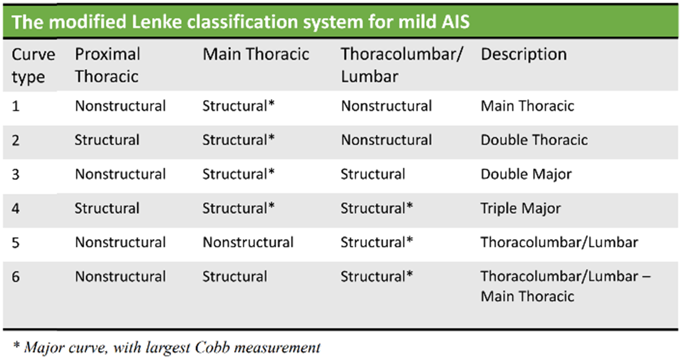
Details of the modified Lenke classification system for mild AIS (m-Lenke)
Key Findings
- 70.1% of the subjects had identical curve classification results and 72.0% had the correct major curve detection compared to X-ray.
- Lumbar-dominated curves had distinctive performance (m-Lenke type 5 with p=0.91, r=0.91).
Citation
– Monitoring of Curve Progression in Patients with Adolescent Idiopathic Scoliosis Using 3-D Ultrasound (2024)
In this retrospective cohort study, 136 adolescents with idiopathic scoliosis (Cobb 10°–85°) underwent paired low‑dose biplanar X‑ray and 3‑D ultrasound (Scolioscan) scans at baseline and again 4–32 months later. Curve progression was defined as a ≥ 5° increase in Cobb angle (radiograph) or Ultrasound Curve Angle (UCA). Both modalities used the same standing posture, ultrasound scans were obtained by sweeping the tracked probe from L5 up to T1.

(a) Participant being scanned with the ultrasound probe (b) Software interface shown during scanning (c) Ultrasound projection images in the coronal plane
Key Findings
- Sensitivity: 0.93 for detecting progression (true positives: 19/21).
- Specificity: 0.90 for ruling out non‑progression (true negatives: 107/115).
- Negative likelihood ratio: 0.08.
- Robust across subgroups: performance stable by age (< 13, 13–15, > 15 y), gender, and initial curve magnitude.
Citation
Evaluation
– 3D ultrasound imaging provides reliable angle measurement with validity comparable to X-ray in patients with adolescent idiopathic scoliosis (2021)
The Scolioscan system mainly consists of a linear ultrasound probe (central frequency of 7.5 MHz and 7.5 cm width) with an electromagnetic spatial sensor attached to it to detect the corresponding position and orientation of the probe. 3D image reconstruction method creates coronal images with more lateral features for angle measurement. This study evaluated the reliability of the angle obtained by Scolioscan and its validity compared to the gold standard Cobb angle measurements from EOS radiography in adolescent idiopathic scoliosis (AIS) patients. Patients were requested to receive both ultrasound scanning and EOS X-ray imaging on the same day.
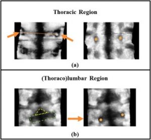
Illustration of placement points for acquiring the measurements
Key Findings
Reliability of Scolioscan angle measurement
- Excellent intra-rater and intra-operator reliability with ICC ≥ 0.973
- Excellent inter-rater and inter-operator reliability with ICC ≥ 0.925
- Overall Mean absolute difference (MAD) ≤ 3.5°; Standard error of measurement (SEM) ≤ 1.7°
Validity compared to EOS Cobb angle
- Very good correlations between Scolioscan angle and Cobb angle for main thoracic (R2=0.893) and (thoraco)lumbar (R2=0.884) curves
- Excellent inter-method variations (ICC ≥ 0.933) with overall MAD ≤ 3.0° and SEM ≤ 1.5°
Citation
– A reliability and validity study for different coronal angles using ultrasound imaging in adolescent idiopathic scoliosis (2018)
In ultrasound imaging of the spine, both the spinous processes (SPs) and transverse processes (TPs) were used to measure the coronal deformity. Both landmarks provided reliable information on the severity of the curve as related to the traditional Cobb angle. However, it remained unclear which coronal ultrasound angle is the most appropriate method to measure the curve severity. This study investigated the reliability and the validity of three Scolioscan angle measurement methods (1. Automatic SP angle; 2. Manual SP angle; 3. Manual TP angle) in the coronal plane as compared with the radiographic coronal Cobb angle in patients with AIS.
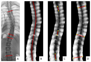
Radiographic coronal Cobb angle (A); Automatic SP angle (B); Manual SP angle (C); Manual TP angle (D)
Key Findings
- Excellent intra- and inter- reliability of all three measurement methods (ICC=0.84-1.00)
- All three coronal ultrasound angles showed excellent linear correlations with the Cobb angles (R2=0.970-0.992)
Citation
– A reliability and validity study for Scolioscan: a radiation-free scoliosis assessment system using 3D ultrasound imaging (2016)
The aim of this study was to test the reliability of spine deformity measurement of Scolioscan and its validity compared to the gold standard Cobb angle measurements from radiography in adolescent idiopathic scoliosis (AIS) patients. The study investigated (1) Intra- and inter- reliability between two operators for acquiring images using Scolioscan and among three raters for measuring spinal curves from those images; (2) Correlation between the Cobb angle obtained from standing plain radiography (within three months before Scolioscan assessment) and the spine curve angle obtained using Scolioscan. Spinal curve angle measurement of Scolioscan was based on the location of spinous processes, as represented by the mid-line of volume projection images obtained.
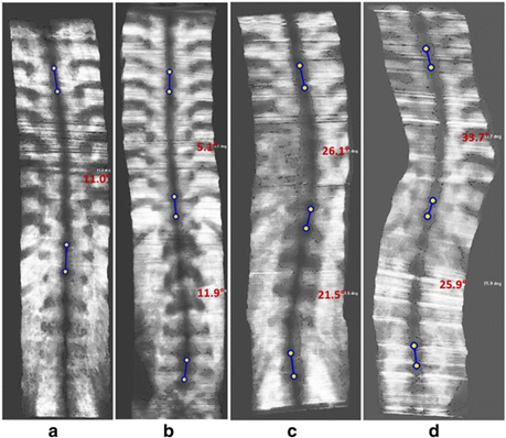
Typical volume projection images obtained and measurements according to spinous processes
Key Findings
- Very good intra-rater and intra-operator reliability with ICC ranging 0.94-0.99 and 0.88-0.97 respectively.
- Very good inter-rater and inter-operator reliability with ICC ranging 0.88-0.93 and 0.87-0.94 respectively.
Citation
– Comparison of two ultrasound methods from Scolioscan® for measuring spinal curvature: spinous processes (SP) versus transverse processes (TP) (2025)
This study compared the validity and reliability of two Scolioscan® automatic ultrasound measurement methods based on the spinous processes (SP) and the transverse processes (TP) against the gold‑standard Cobb angle from standing anterior–posterior X‑rays in 71 adolescent idiopathic scoliosis patients. All patients underwent both X‑ray and ultrasound assessment on the same day. SP-based angles were multiplied by region‑specific conversion factors (1.43 for thoracic, 1.23 for lumbar), whereas TP‑based measurements required no conversion.
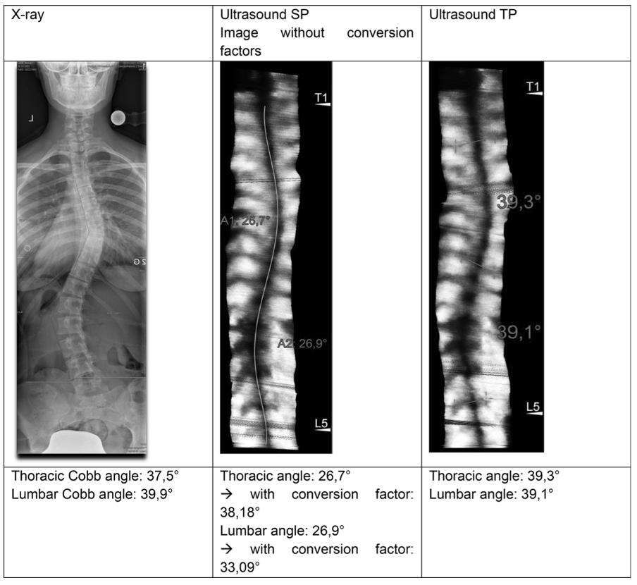
Example of a patient with the according X-ray image, ultrasound images based on SP and TP
Key Findings
- SP method: Thoracic ICCs ranged from 0.871–0.900. Lumbar ICCs ranged from 0.702–0.799.
- TP method: Thoracic ICC = 0.893 (95% CI 0.834–0.932). Lumbar ICC = 0.888 (95% CI 0.823–0.929).
- Both methods achieved ≥ good reliability, with TP showing marginally higher and more consistent ICCs, especially in the lumbar region.
Citation
Technology
– Volume reconstruction of freehand three-dimensional ultrasound using median filters (2008)
3D ultrasound imaging has proven to be able to overcome the limitations of 2D ultrasound when visualising and analysing 3D anatomies. Freehand imaging with spatial tracking devices was developed as one of the methods to obtain 3D images, where the ultrasound probe is moved by hand in an arbitrary manner to obtain a sequence of 2D images (B-scans). This study aims to apply median filters during reconstruction of B-scans into 3D data set for reducing interpolation error and improving the quality of 3D images in a freehand 3D ultrasound system. Four algorithms including the standard median (SM), Gaussian weighted median (GWM) and two types of distance-weighted median (DWM) filters were proposed to filter noises and compute voxel intensities.
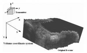
B-scans transformed to the volume coordination system for reconstruction
Key Findings
- Compared with the voxel nearest-neighbourhood (VNN) and distance-weighted (DW) interpolation methods, the four median filters reduced the interpolation error by 8.0–24.0% and 1.2–21.8%, respectively, when 1/4 to 5 B-scans was removed from the raw B-scan sequence.
Citation
– Automatic Measurement of Spine Curvature on 3-D Ultrasound Volume Projection Image With Phase Features (2017)
An automated measurement of spine curvature in ultrasound volume projection imaging (VPI) was proposed, using prior knowledge in vertebral anatomical structures. It is based on the extraction of bony features from VPI images using a newly proposed two-fold thresholding strategy, with information of the symmetric and asymmetric measures obtained from phase congruency. The spinous column profile is detected from the segmented bony regions, and it is further used to extract a curve representing spine profile. The spine curvature is then automatically calculated according to the inflection points along the curve. The algorithm was evaluated on volunteers with the different severity of scoliosis.
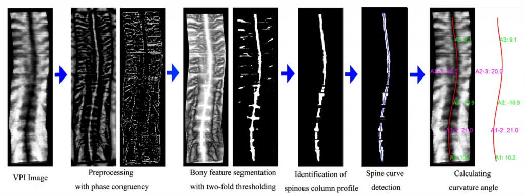
Flow diagram of the automatic algorithm
Key Findings
- Significant linear correlation between the spinous process angle measured manually and the automatic method (slope = 0.86, r = 0.90). Mean difference is 2.6 degrees.
- Significant linear correlation with X-ray Cobb angle (slope = 0.94, r = 0.83)
Citation
– Automating spine curvature measurement in volumetric ultrasound via adaptive phase features (2019)
In previous research, the automatic methods of spine curve detection in volume projection imaging (VPI) images were proposed. However, there were limitations such as degenerating accuracy due to other bony features. The new automatic method is based on prior knowledge about the geometric arrangement of the spinous processes and overcomes previous limitations. The frequency bandwidth of log-Gabor filters is adaptively adjusted to calculate the oriented phase congruency, facilitating the segmentation of the spinous column profile. And the spine curvature angle is finally calculated according to the inflection points of the curve over the segmented spinous column profile. The performance of the automatic method is evaluated on spine VPI images among patients with different scoliotic angles.
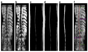
(a) Original VPI-SP image. (b) The enhancement using histogram equalization. (c) The phase congruency using log-Gabor with σf = 0.1. (d) The phase congruency using log-Gabor with σf = 0.15. (e) The segmentation of spinous column profile. (f) The final result of spine curve detection and spine curvature measurement.
Key Findings
- Curvature angles obtained by the automatic method have a high linear correlation with those by the manual method (r = 0.90, p < 0.001) and X-ray Cobb’s method (r = 0.87, p < 0.001)
Citation
– Ultrasound Sagittal Projection Imaging for the Assessment of Scoliosis (2020)
To obtain both coronal and sagittal radiographic images for comprehensive scoliosis assessment, 2 radiographs have to be taken. A novel projection imaging method (SPI) was developed to obtain both coronal and sagittal projection images of the spine from a single ultrasound volume data set. The method started with deriving spine curves from coronal images at different depths and constructed a non-plane surface, followed by rendering the sagittal spine visualization using the average blending function. Its performance was evaluated on 30 subjects who were requested to take X-ray to obtain sagittal images for Cobb angle measurement.
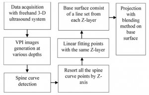
Flow diagram of the sagittal projection image generation
Key Findings
- The spine anatomy in ultrasound projection images are well consistent with those presented in the X-ray radiograph on both coronal and sagittal planes
- High linear correlation between the spinal curvature measurements from SPI and radiographs (slope = 0.93, r = 0.85, p < 0.001)
Citation