

There are no special qualification requirements for users. Users can operate the products after attending the short-term training held by Telefield Medical Imaging Limited, and according to the relevant requirements of the user manual. With the continuous improvement of the functions, we suggest users to participate in advanced training arranged by manufacturer / authorized service providers from time to time.
It takes about 30 seconds only for one scan and costs about 15 minutes for a whole assessment with one-to-one service, including preparation, clothes changing, angle measurement and report explanation. During mass screening program, a pipeline approach, with different tasks supported by different staff, can be used to reduce the time for each subject down to 3-5 mins.
The suggested range of height is 1 to 2 meters
Scolioscan® is applicable for people of all ages. So far, the results we have collected for the youngest kid was 2 years old.
The use of ultrasound gel is to allow the penetration of ultrasound waves to our body.
If ultrasound gel is missing, we cannot perform the scanning as the ultrasound waves cannot pass through the body. This is a normal practice in any ultrasound examination.
Yes. It can scan the patients in any postures. For the patients in supine position, a hollow slot aligns with the spine is necessary in order to expose the back for scanning.
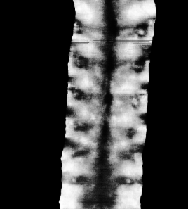
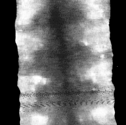
The operator first applies ultrasound gel on the back of subject, sets a scanning range according to the height of subject, then performs the scan on the spine covering the 5th lumbar vertebra (L5) to the 1st thoracic vertebra (T1) from bottom to the top. There will be a real-time preview image of the spine along the coronal plane during the scan for reference. The whole scan can normally be completed within 30 seconds.
After the scanning, a coronal image of the spine can be obtained. Measurement of spine deformity can be done manually or automatically for analysis. There are two different ways for the measurement using different spinal features.
We recommend using UCA to perform angle measurement.
The drawing line standard for determining the points on both sides of the upper and lower vertebrae to represent the most inclined vertebrae is as follows:
- The most inclined vertebrae
- The determination of the locations of the clearest, most symmetric, and most informative points can refer to the following order
| A. Thoracic image
1) White dots in the black region bilaterally |
 |
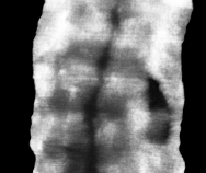
| 2) If the white dots are relatively small and the midpoint of the black region can easily be recognized, the midpoint of the black region is taken bilaterally. | 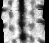 |
| 3) If the bilateral dark marks difficult to identify, the bilateral symmetric white areas can be used. In this case, the highest points of the white area on both sides are used (in other cases, the centers, bottom, etc. of bilateral while area can be used). | 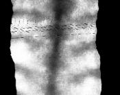 |
| B. Lumbar image
4) The straight edges across the black shadow on both sides 5) Bilateral symmetrical black shadow edges 6) Bilateral symmetrical black shadow |
|
| 7) If the above information is difficult to obtain, make a vertical line for the most inclined part of the black midline (shadow of spinous process) | 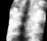 |
Yes, it is possible. Scolioscan® can be applied to assess the spinal flexibility for surgical planning, prognostic outcome, follow-up with post-operational patients, etc. Besides, the scanned image can be analyzed using the 3D analysis software, Scolio3D. Some metal implant may result in interference of the image quality, but patients can still undergo the scanning.
Yes, it is possible. Scolioscan® can be applied to assess the cervical vertebrae. However, the contact between the probe and the skin surface may not be satisfying to collect comprehensive information due to the contour of the cervical region. Please note that this application has not been registered yet at this moment, while it can be used.
Yes, Scolioscan® can assist in the design, fitting and follow-up of the brace treatment. These can benefit from the unlimited scanning frequency due to its radiation-free feature. Besides, with the 3D analysis software Scolio3D, more information can be accessed for analysis purpose, including in sagittal and transverse planes.
During design:
Baseline scoliotic curve for comparison purpose. Spinal flexibility in prone, supine, side bending, forward bending positions can be collected for bracing strategies1.
Real-time feedback can be provided by adjusting the location, orientation and force of the external paddings to mimic the in-brace correcting effect.
During fitting:
Real-time feedback can be provided as a reference to adjust the paddings, straps tightening.
Follow-up:
No restrictions on scanning frequency that in-brace correction and curve progression can be closely monitored. Brace treatment strategy can be adjusted timely.
1 He, C., & Wong, M. S. (2018). Spinal flexibility assessment on the patients with adolescent idiopathic scoliosis: a literature review. Spine, 43(4), E250-E258.
Yes, as long as it can have a good contact of the probe and skin surface on the back. There are mainly 3 suggestions:
- If the brace is designed with back opening, it can be scanned along with the opening slot.
- If the brace is designed with back opening, and there are some straps blocking the scanning path, we can use a grip to grab the brace on the lateral side of the brace to estimate the correcting effect after tightening the straps. And to release the straps for the scanning.
- If the brace is designed with front opening, there is one published literature indicating that the scoliotic spine would gradually deform to the original curvature from in-brace situation in 2 hours. Theoretically, after the patient wearing the brace for more than 2 hours, we conduct the scanning right after the patients taking off the brace may represent the in-brace situation2
2. Li, M., Wong, M.S., Luk, K.D., Wong, K.W. and Cheung, K.M., 2014. Time-dependent response of scoliotic curvature to orthotic intervention: when should a radiograph be obtained after putting on or taking off a spinal orthosis?. Spine, 39(17), pp.1408-1416.
Yes, Scolioscan® can generate a one-page report including an image in coronal view together with measured angle. This report can be printed out with remarks. Besides, the image in the sagittal plane information can be shown in the 3D analysis software Scolio3D.
Yes, it allows scanning on different body parts, including shoulder, pelvis, etc. We can utilize 3D analysis software Scolio3D to display the shoulder and pelvis. Scolio3D can provide 3D view for different body parts scanned.
Patients with below symptoms or conditions should not use Scolioscan®:
- Implanted with pacemakers, pain modulators, insulin delivery systems, cochlear and any defibrillator
- Ferromagnetic implants
- Weighed over 150kg