

“Scolioscan® can realize the correction and visualization of posture (standing, sitting), so their medical experiences can be supported by medical science. The device can monitor the patients with small degrees of scoliosis, and provide reference to fit, review and adjust the brace. This device can also provide scientific evidence for postural correction exercises, weight-bearing changes, and body alignment for scoliosis patients.”
——Associate Professor Zhang Xiaohui The Department of Sports Medicine of Guangzhou Sports University
About Department of Sports Medicine, Guangzhou Sport University
The scoliosis project of the Department of Sports Medicine of Guangzhou Sports University has successively received funding from Provincial Innovation Colleague and Guangdong Provincial Science and Technology Project, and has established cooperation with Coventry University, the Hong Kong Polytechnic University and many scientific research institutions. They are holding various volunteer medical consultation and intensive training in Guangzhou, and more than 1,200 patients with mild and moderate scoliosis are diagnosed and treated each year, with good treatment outcome.
Source: Guangzhou Sport University
The scoliosis project of the Department of Sports Medicine of Guangzhou Sports University is led and created by Professor Liao Bagen, the leader and doctoral supervisor of the Department of Sports Medicine of Guangzhou Sports University, Associate Professor Zhang Xiaohui and others. With the experience and achievement from different conservative treatment, from research to clinical, the team keeps exploring and solving the tough problems on scoliosis correction.
This project has successively received funding from Provincial Innovation Colleague of Innovation and Guangdong Provincial Science and Technology Project, and has established cooperation with Coventry University, the Hong Kong Polytechnic University and many scientific research institutions. They are holding various volunteer medical consultation and intensive training in Guangzhou, mild and moderate scoliosis are diagnosed and treated with good treatment outcome.
“Scolioscan® is the world’s first three-dimensional ultrasound system with application on scoliosis examination, providing a non-invasive and radiation-free examination method for scoliosis patients. Using three-dimensional ultrasound to assess coronal deformity of adolescent idiopathic scoliosis is more reliable, and it is good for screening, progress monitoring and evaluation on treatment outcomes.
——Professor Zheng’s team has verified the reliability of Scolioscan®, and the article was published in “Scoliosis and Spinal Disorders”. In 2018, Scolioscan® is installed in the Guangzhou Sport University, and carried out the evaluation program using non-invasive and radiation-free 3D ultrasound in the summer of that year.
Their Pain Point and our Solution
(1) Postural Correction Visualization by Using Scolioscan®
The Department of Sports Medicine of Guangzhou Sports University makes use of the Scolioscan® Ultrasonic Scoliosis Assessment System to evaluate the correction effect of scoliosis patients: the correction of upper thoracic curvature after wearing a brace; the weight-bearing examination was carried out for the cases of Rigo A and B scoliosis patients with obvious body deviation. It is verified that the designed movement exercises make the spine tend to the midline, which provides theoretical support on the formulation of exercise plan.
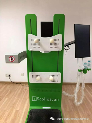
Figure 1. Scolioscan® Ultrasound Scoliosis Assessment System was installed in the Department of Sports Medicine of Guangzhou Sports University
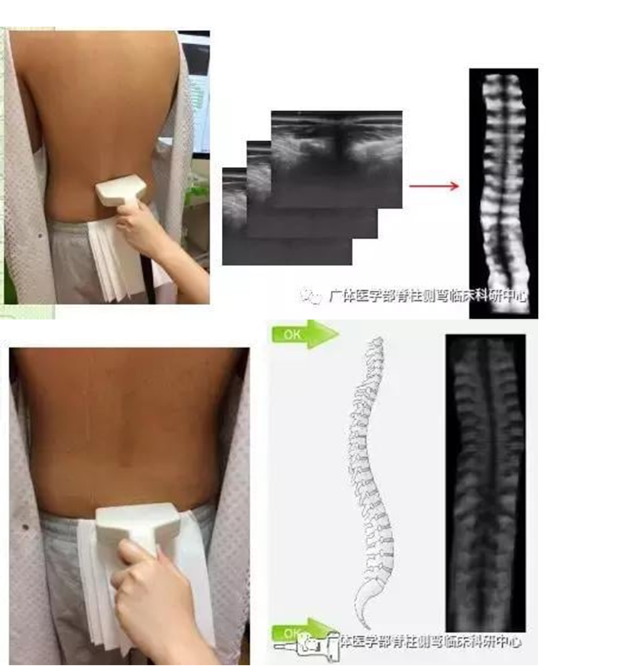
Figure 2. Scoliosis clinical research center of the Department of Sports Medicine of Guangzhou Sports University undergoes correction evaluation
(2) Cases of Posture Correction Visualization
- Case (1)
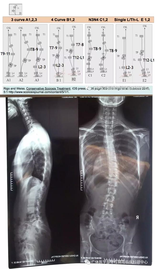
Figure 3. According to the X-ray image: Type RigoA1, Cobb angle 26°to the right in T5-L3
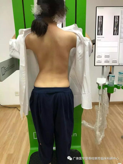
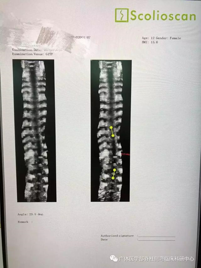
Figure 4. In natural standing posture, the measured angle using 3D ultrasound is 24°, and the error is 2° varied from the measurement of the X-ray image
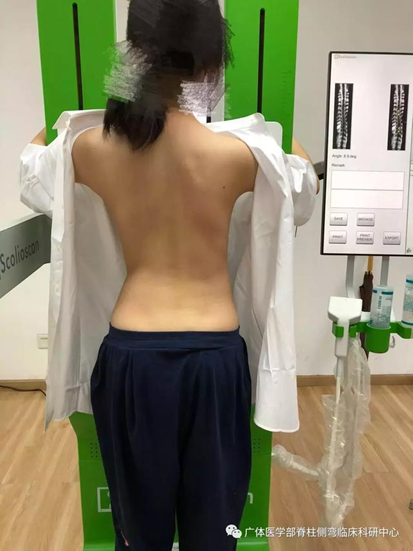
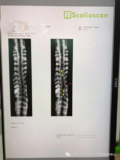
Figure 5. After instructing the patient’s left leg with weight bearing and correcting the standing posture, the measured angle is 9° using 3D ultrasound.
- Case (2)
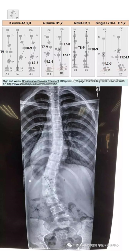
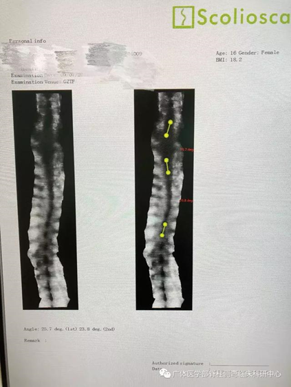
Figure 6. According to X-ray image: Type RigoB1, Cobb angle 22°to the right in T4-T10, Cobb angle 42°to the left in T10-L4
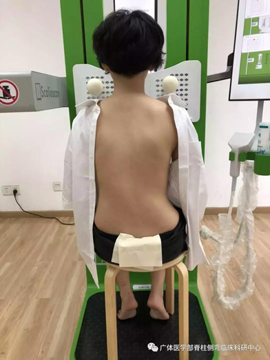
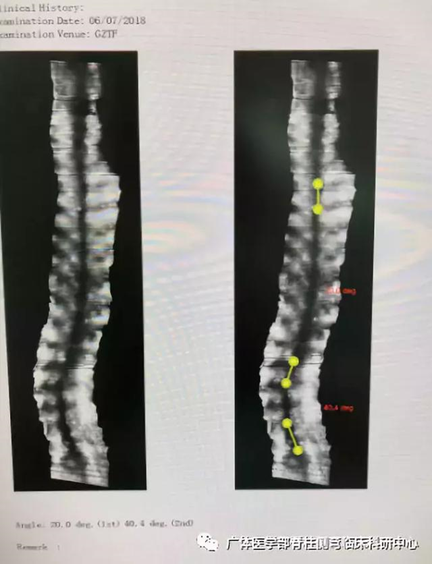
Figure 7: In natural sitting posture, the measured angle using Scolioscan 3D ultrasound is 20°to the right in T4-T10, 40°to the left in T10-L4 (the error is 2° varied from the measurement of the standing X-ray image)
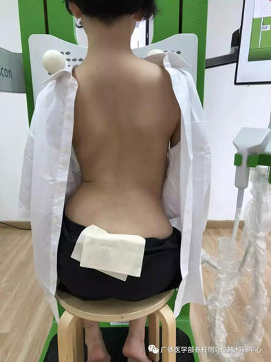
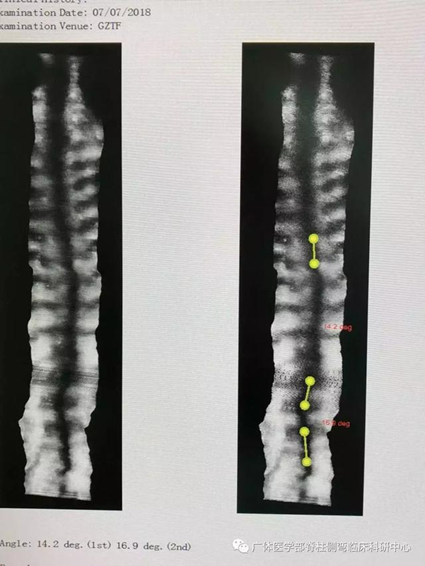
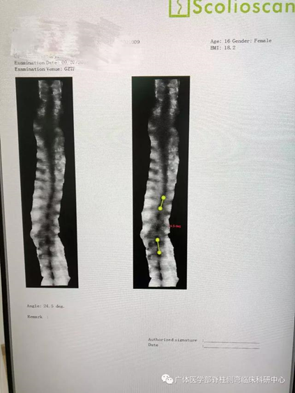
Figure 8: After instructing the patient’s right hip with weight bearing and correcting the posture, the measured angle is 14°to the right in T4-T10 and 17°to the left in T10-L4 using 3D ultrasound.
- Case (3) ——thoracic curve
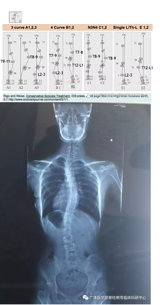
Figure 9. According to X-ray image: Type RigoB1 (with thoracic curve), Cobb angle 30°to the left in T1-T4, Cobb angle 29°to the right in T5-T11, Cobb angle 28°to the left in T11-L3
In natural sitting posture, the measured angle using Scolioscan 3D ultrasound is 26°to the left in T1-T4, 24°to the right in T5-T11, 25°to the left in T11-L3 (the error is 5° varied from the measurement of the standing X-ray image) After instructing the patient to practice a specific sitting posture: imitate sitting on the left side of the classroom and look up at the moon on the right to watch the slides, keeping the weight on the right hip and the body trunk straight
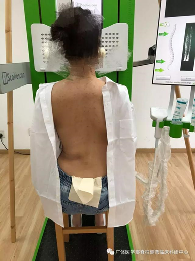
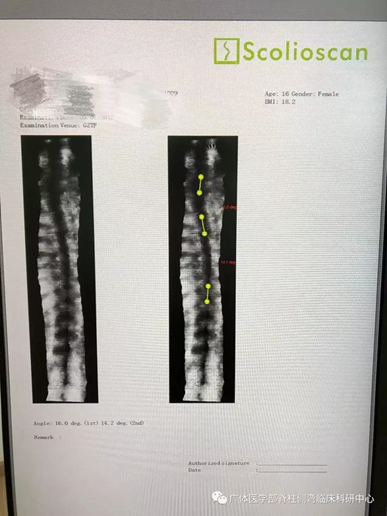
Figure 11. Measured angle by 3D ultrasound: 16°to the left in T1-T4, 14°to the right in T5-T11; T11-L3 was corrected and no degree was measured. Combined with posture correction for thoracic curvature: change the weight bearing, adjust the body line, and maintain the posture of the shoulder balance, which has scientific basis
(3) Scoliosis Intensive Training Camp in summer
Guangzhou Intensive Training Camp has been held since 2018. A total of 40 trainees from inside and outside Guangdong Province, as well as Hong Kong and Macao participated in the training. Among them, college students who studied in Canada and the United Kingdom returned to Guangzhou for participating the training as well. During the training camp, the team has also completed the correction review for more than 200 youngsters with scoliosis.
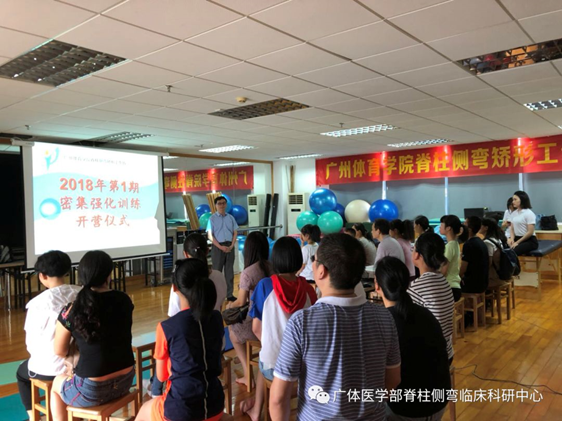
Figure 12: The opening of Guangzhou Intensive Training Camp
In the training camp, the team will classify the children according to their physical appearance, physical fitness, movement patterns and imaging diagnosis, and use Scolioscan® to provide visualized analysis on the children and formulate targeted and personalized plans.
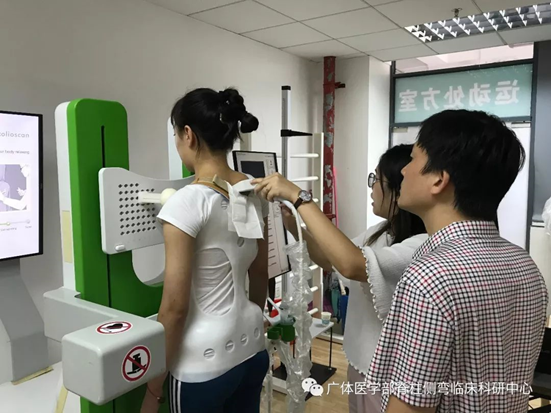
Figure 13. Therapist uses the Scolioscan® assessment system to diagnose and evaluate the correction of using the thoracic brace
During the training camp, Professor Zheng Yongping from the Hong Kong Polytechnic University, the inventor of the Scolioscan® Ultrasound Scoliosis Assessment System, led the team to provide technical guidance on the application of the assessment system. Prof. Zheng appreciated the innovative application of “posture correction visualization” by the scoliosis project of the Department of Sports Medicine of Guangzhou Sports University, and gave valuable suggestions.
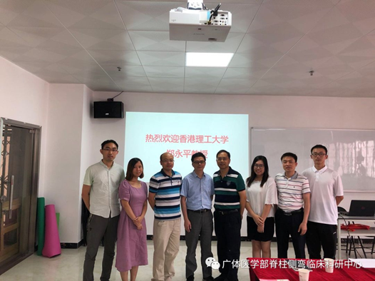
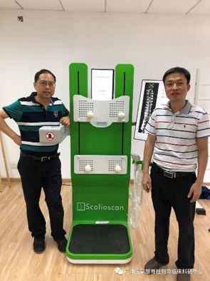
Figure 14. Technical coaching by the Scolioscan® team during the training camp
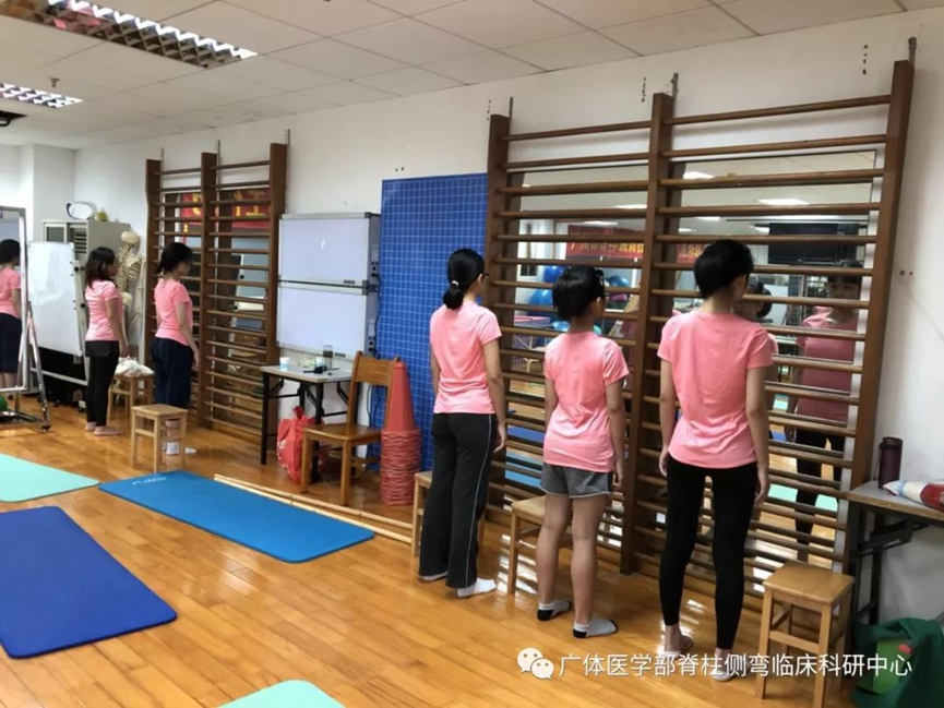
Figure 15. Children learn how to change their standing posture with weight-bearing, which is the basis of postural correction
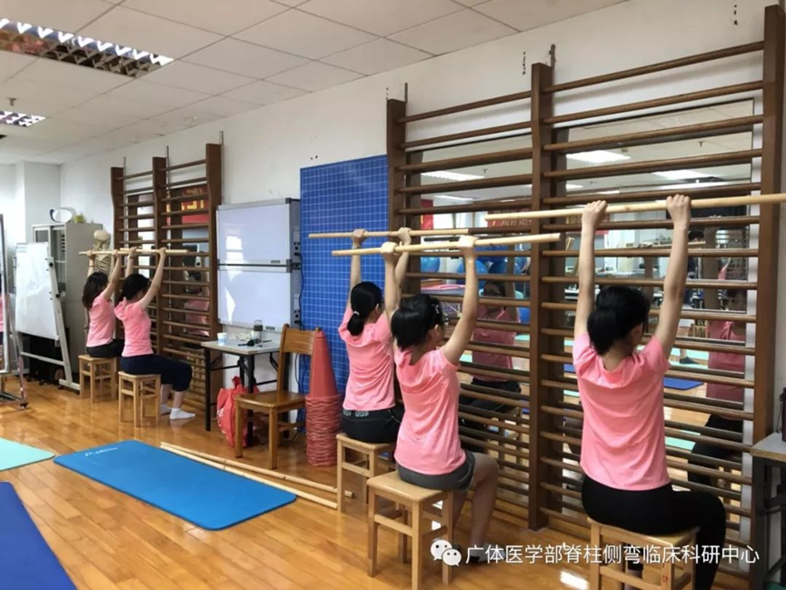
Figure 16. Sitting posture exercises after adjusting the force line
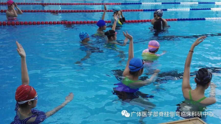
Figure 17. Water resistance exercises are useful for posture correction, using resistance exercises to quickly strengthen the paraspinal muscles that stabilize the spine, in order to facilitate correction
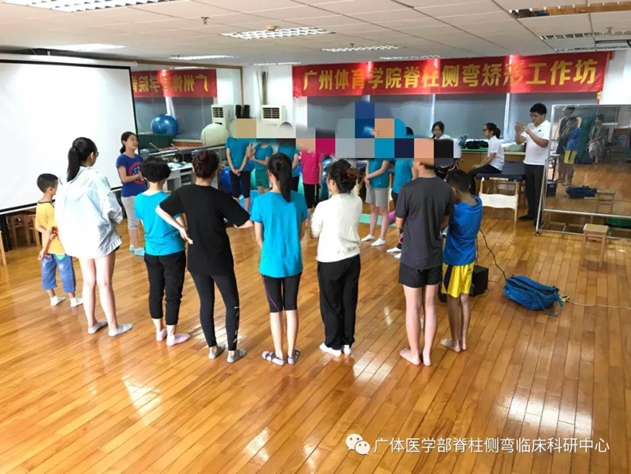
Figure 18. The author of the child book shares the personal experience
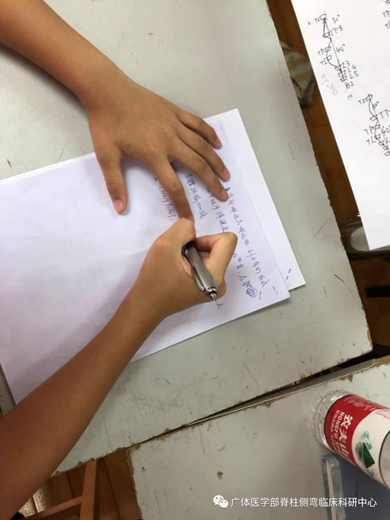
Figure 19. Children is drawing spine curves and writing the outline of exercise prescriptions.
At the closing ceremony, the children and their parents shared their experiences and sang the closing song together. The teacher awarded meaningful souvenirs to the students, and hoped that the children can continue to overcome challenging difficulties, and will receive good news after half a year for review.